A laminoplasty is performed to relieve pressure on the spinal cord. Disc herniations and bone spurs commonly cause increased pressure in the spine. This surgery involves cutting vertebrae in two locations allowing the Orthopedic surgeon to prop the bone open, which in turn decreases pressure and provides the spinal cord with adequate room to heal.
What is it?
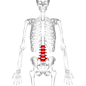
A laminoplasty is a surgical procedure performed to create space between spinal vertebrae, which relieves pressure on the spinal cord and facilitates healing. This procedure is performed on most regions of the spine, such as the cervical, thoracic, or lumbar regions, and the laminoplasty is usually performed through a posterior approach. Arthritis, bone spurs, tumors, fractures, and degenerative changes are all conditions that may cause increased intervertebral pressure. Laminoplasty is also used to treat myelopathy. Patients experiencing severe myelopathy often suffer from intense pain, muscle weakness, spasticity, muscle atrophy, and sensory deficits.
What should I do to prepare?
You should not eat or drink after midnight prior to the surgery. You will need to stop taking certain medications, especially NSAIDs, a week before the procedure. Some doctors may suggest exercising and eating healthier in the weeks before your procedure. There is accumulating evidence suggesting surgeries are more successful when patients are active and maintain a healthy diet. Your surgeon will also strongly recommend you stop smoking weeks before the procedure.
What happens during the process?
Depending on the location of the increased pressure in the spinal cord, your surgeon will make an incision in either the patient’s neck or back. Following this initial incision, the appropriate vertebra is “hinged.” This involves cutting through the lamina on one side and thinning out the bone on the other side of the vertebrae. The thinner side acts as a hinge while the other side is held open with a surgical wedge or piece of bone. Increasing the space inside the spinal column relieves the pressure damaging the spinal cord. Then, the spinal cord begins to heal and move away from the source of the pressure. Sometimes, the surgeon implants a shunt to monitor bleeding following the surgery, and typically removes this device the following day.
What are the risks and potential complications?
As with any surgery, complications and risks are a possibility. Some of these include excessive bleeding and infections, anesthetic complications, pain, numbness, or paralysis due to damage of nerves/spinal cord, bladder or bowel incontinence, cerebrospinal fluid leak, and unresolved pain following the surgery.
Disclaimer:
All GlobeHealer Site content, including graphics, images, logos, and text, among other materials on the site are for educational purposes only. This content is not intended to be a substitute for professional medical advice, and you should always contact your physician or qualified health provider for information regarding your health. Information on this site regarding the overview, diagnosis, and treatment of any kind should be looked at, in addition to the advice and information of your health care professional. Do not disregard medical advice or delay seeking treatment or medical advice due to information found on the GlobeHealer site.
If there is even the possibility that you may have a medical emergency, seek treatment, call your doctor, or call your local emergency telephone number immediately. GlobeHealer does not endorse being the first line of communication in case of emergency and does not endorse any specific test, physician, facility, product, procedure, opinion, or other information that is or may be mentioned on this site or affiliated entities. Reliance of any and all information provided by GlobeHealer, its employees, affiliations, others appearing on the Site under the invitation of GlobeHealer, or visitors of the site is solely at your own risk and is not the responsibility of GlobeHealer.
Image Source: https://upload.wikimedia.org/wikipedia/commons/5/53/Lumbar_vertebrae_anterior.png